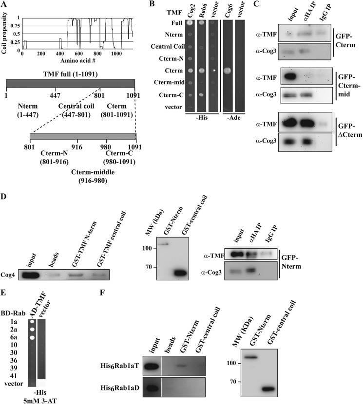
FIGURE 3.
Mapping of COG interaction sites on TMF. A, coil propensity of TMF (16, 59), and the derived constructs representing various regions of the golgin. B, Y2H interactions of TMF fragment prey constructs with Cog2, Cog6, and Rab6 baits. C, co-immunoprecipitation of TMF fragments with the COG complex. Experiments as in Fig. 2B using the GFP tagged TMF constructs Cterm, Cterm-mid, and ΔCterm expressed in Cog1HA cells. Inputs are 0.1% of precipitate for TMF and 3% for Cog3. D, probing the interaction between COG and TMF Nterm. Partially purified COG (as in Fig. 1C) was pulled down with GST tagged Nterm and central coil fragments. COG was probed by anti-Cog4 immunoreactivity (first column). The amounts of used TMF fragments probed with anti-GST are shown in the middle panel. GFP-TMF N-term expressed in Cog1HA cells was immunoprecipitated as in C (bottom panel). E, yeast two-hybrid interactions between full-length TMF prey fusions and active (T) Rabs. F, GST-TMF fragments were used to pull-down recombinant His-tagged Rab1a as in D. GDP and GTP locked mutants of Rab1a loaded with the appropriate nucleotide were used. The Rab1aT and Rab1aD images are from the same blot exposed for the same time, but have been rearranged in the figure for more clarity. TMF fragment levels are shown on the right as in D.

