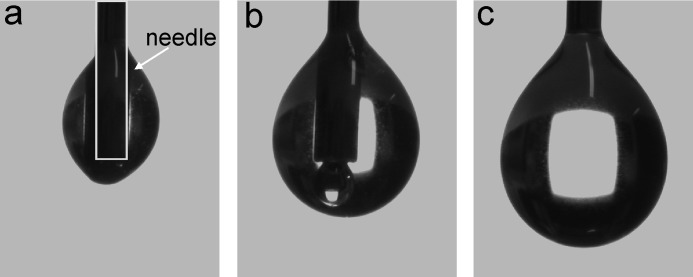
FIGURE 3.
Adherence of EPL1 solutions to needles. The drop dosing system and the high focus camera integrated in the DSA 100 contact angle device allow the imaging of small droplets with defined volume on the outlet needle. The formation of 0.5-μl droplets of an EPL1 solution is shown. Instead of hanging at the outlet of the needle as is usually observed for aqueous solutions, EPL1 crawls up along the needle (a and b) and only forms a hanging droplet when the gravitational force is strong enough to pull the droplet down (c). The shape of the needle is indicated in a with a gray box.

