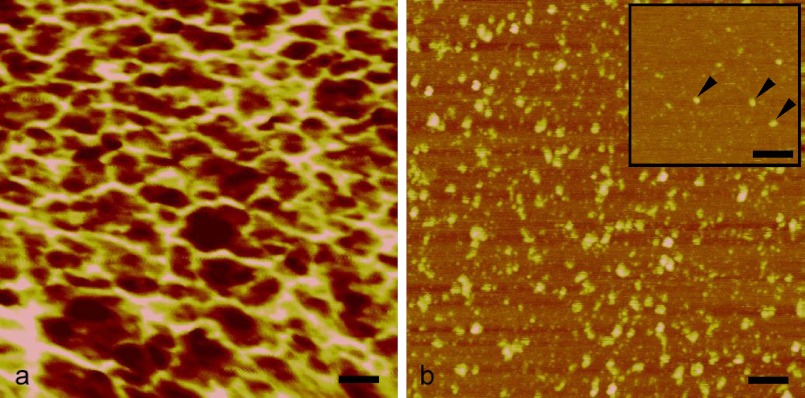
FIGURE 5.
AFM analysis of EPL1. a, at a concentration of 0.5 mg/ml, protein molecules assemble into an irregular meshwork. b, even at concentrations of 1 μg/ml, an agglomeration can be observed. Isolated single molecules were detected only at concentrations as low as 0.1 μg/ml (arrows in inset). Protein monomers shown in the inset have a diameter of approximately 4 Å, which is close to the approximate dimensions of CP (Protein Data Bank code 2KQA), based on its three-dimensional structure. Scale bars, 50 nm; height scale, 4 nm from dark to bright.

