
FIGURE 9.
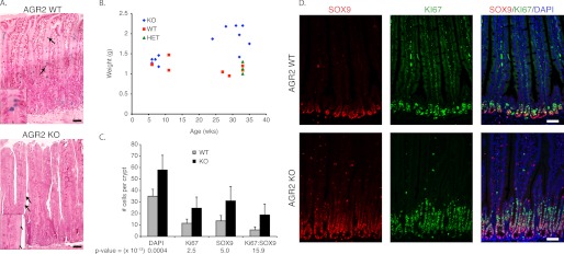
The small intestine of Agr2 KO mice exhibit decreased mucin and increased weight, cell proliferation, and nuclear SOX9. A, hematoxylin and eosin stained the small intestine of Agr2 WT and KO mice. Mucus-secreting goblet cells are stained with Alcian blue. The arrows highlight Alcian blue-stained goblet cells, and a magnified view is displayed in the inset. B, shown is a scatter plot of mouse age versus small intestine weight of Agr2 KO (KO), WT (WT), and heterozygotes (HET). C, shown is the average number of SOX9- and Ki-67-labeled cells per crypt (WT, n = 41 crypts; KO, n = 51). p values ( × 10−13) between WT and KO are listed below each measured label. Error bars represent ± 1 S.D. D, double-labeled immunofluorescence for SOX9 (red) and Ki-67 (green) is shown. The nuclei are stained blue with DAPI. The scale bar in all images represents 50 μm.