
FIGURE 5.
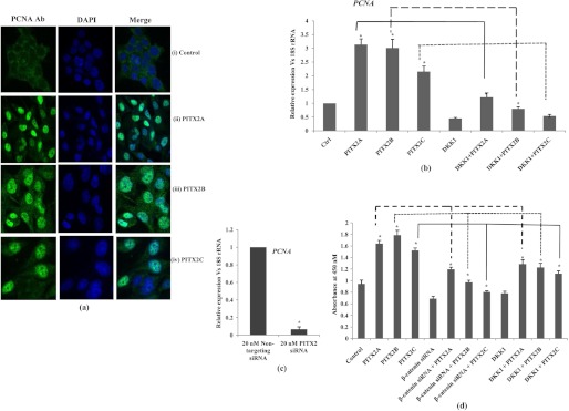
PITX2 enhances proliferation in SKOV-3 cells. a, confocal staining for PCNA is performed in synchronized cells transiently transfected with either empty vector (panels i), PITX2A (panels ii), PITX2B (panels iii), or PITX2C (panels iv) expression vectors. The left panels show the images of cells stained with anti-PCNA antibody followed by anti-rabbit Alexa Fluor-488 (green). The nuclei are stained with DAPI (middle panels), and the right panels show the merged image. The images are taken at same exposure time. Scale bar, 20 μm. b, relative expression of PCNA is measured by Q-PCR in the RNAs extracted from PITX2-overexpressed cells and from cells treated either with human recombinant DKK1 protein alone or in combination with PITX2A, PITX2B, and PITX2C expression vectors. c, Q-PCR is performed with the RNA of nontargeting and PITX2 siRNA-transfected cells using primers of PCNA. The comparative expression of the respective gene is shown as relative fold change in the y axis (mean ± S.E.). d, cell growth is assessed by BrdU incorporation assay after transient transfections of PITX2A, -B, and -C or in combination with β-catenin siRNA or DKK1. * represents p < 0.05.