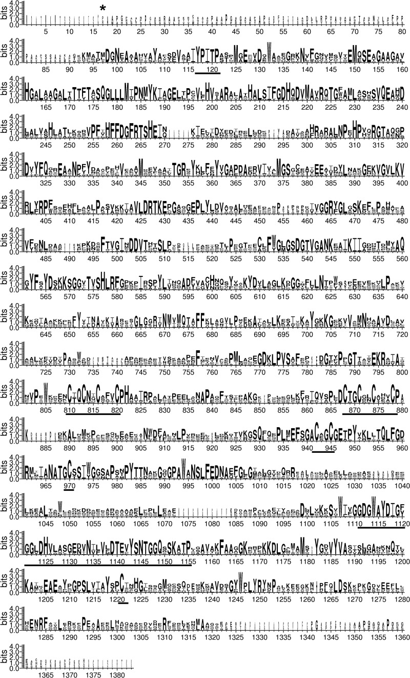
FIGURE 2.
Stacked polypeptide alignment of pyruvate:ferredoxin oxidoreductase primary sequences. Eight PFOR sequences were used for an alignment using ClustalW2 and WebLogo 3 (89, 90). These sequences were C. reinhardtii PFR1 derived from the cDNA obtained in this study, Volvox carteri f. nagariensis (Phytozome v8.0, V. carteri Vocar20008508m), Chlorella variabilis (GenBankTM EFN55341.1), C. acetobutylicum PFO (GenBankTM NP_348846.1), Clostridium pasteurianum (GenBankTM AAD55756.1), Desulfovibrio africanus POR (GenBankTM CAA70873.1), E. coli YdbK (GenBankTM YP_002999180.1), and Synechococcus sp. PCC.7002 NifJ (GenBankTM ACA99434.1). The conserved YPITP substrate-binding site (58), as well as three [4Fe-4S]-cluster coordinating motifs and the region homologous to TPP-binding (59, 91) sites, are underlined. Note that the third [4Fe-4S]-cluster-binding site is atypical and consists of the CXXC motif at positions 942–945 and two separated cysteines at positions 970 and 1221 (60, 61). The first amino acid of recombinant PFR1 is marked by an asterisk.

