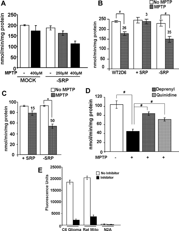
FIGURE 6.
Inhibition of mitochondrial complex I (NADH oxidoreductase) activity following exposure to MPTP. A and B, complex I activity was measured in mitochondria isolated from undifferentiated Neuro-2A cells (50 μg of protein each) treated with or without MPTP. Numbers over the bar diagram in B show % inhibition by added MPTP compared with the nontreated control cells. C, complex I activity was measured in mitochondria from differentiated Neuro-2A cells expressing different CYP2D6 cDNAs. Assays were run as in A. Effects of the CYP2D6-selective inhibitor quinidine (10 μm) and the MAO-B inhibitor deprenyl (10 μm) on complex I activity in mitochondria of −SRP cells were tested. D, inhibition of complex I in cells expressing −SRP 2D6. E, MAO activity was measured in mitochondria from rat liver, C6 glioma, and Neuro-2A cells using Amplex® Red monoamine oxidase assay kit, as per the manufacturer's protocol. Fluorometric assay of MAO-B was carried out using benzylamine as the substrate, based on the extent of inhibition by MAO-B inhibitor pargyline (10 μm). The activity was measured with 530 nm excitation and 590 nm emission. Results represent mean ± S.E. from three separate assays. * denotes p < 0.05 and # denotes p < 0.001.

