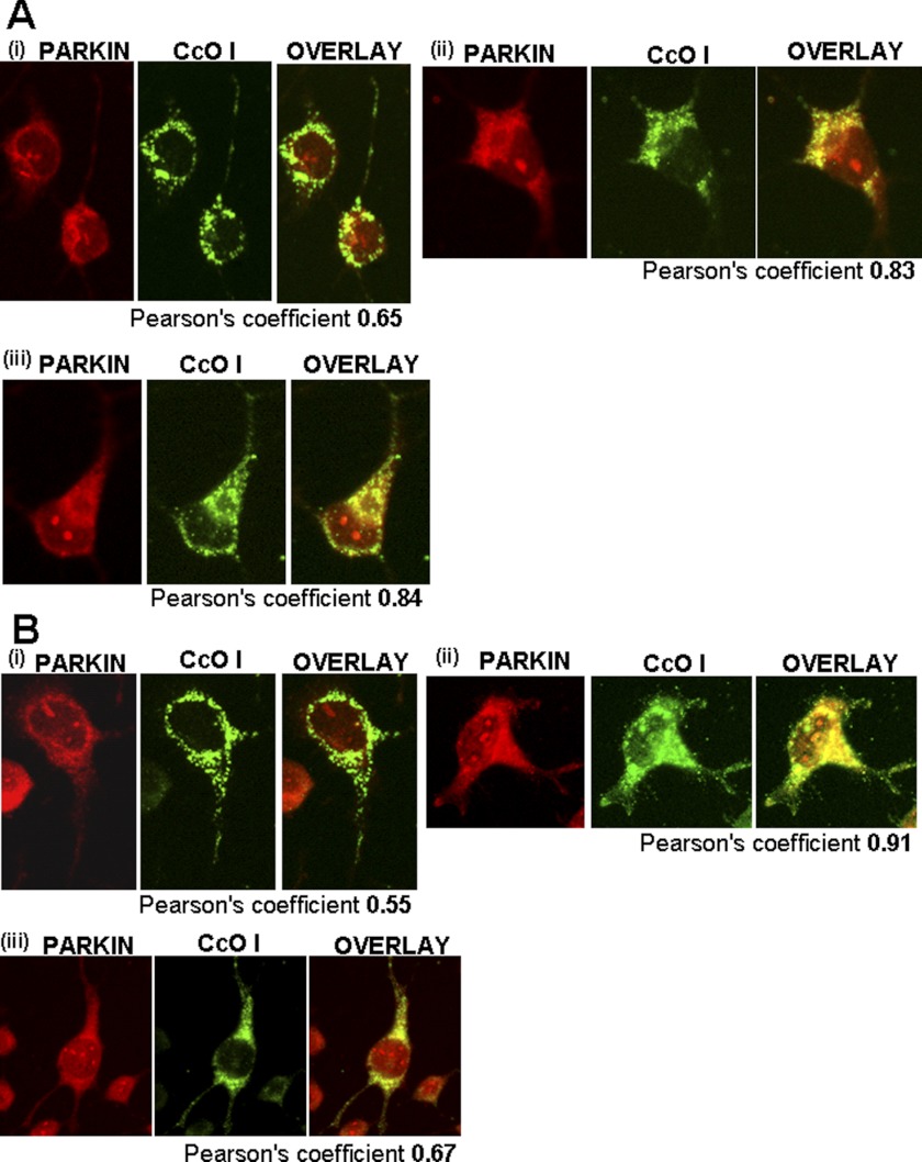
FIGURE 8.
Effects of MPTP on mitochondrial localization of autophagy marker, Parkin. Immunofluorescence microscopy was carried out in differentiated Neuro-2A (Mock and −SRP cells) with and without added MPTP (400 μm) for 48 h and the CYP2D6-selective inhibitor quinidine (10 μm). Cells were incubated with a 1:1000 dilution (v/v) of primary anti-rabbit antibody to Parkin (Abcam, Cambridge, MA) and co-stained with a 1:500 dilution (v/v) of cytochrome oxidase I (anti-mouse) antibody as a mitochondrion-specific marker (Abcam, Cambridge, MA). The cells were subsequently incubated with Alexa 546-conjugated anti-rabbit and Alexa 488-conjugated anti-mouse IgG for colocalization of fluorescence signals. A, mock-transfected cells; B, −SRP 2D6-expressing cells. Panel i, cells with no MPTP treatment; panel ii, cells with added MPTP; panel iii, cells with added MPTP and quinidine. Numbers indicate Pearson coefficients calculated using Volocity 5.3 software.

