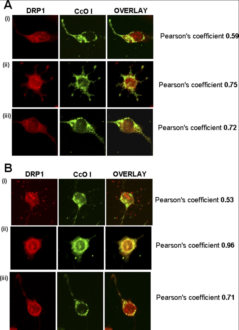
FIGURE 9.
Effects of MPTP on the induction of Drp-1, a marker for mitochondrial fission. Immunofluorescence microscopy was carried out in differentiated Neuro-2A (mock and −SRP cells) with and without MPTP (400 μm) for 48 h as described in Fig. 7. Cells were incubated with a 1:250 dilution (v/v) of anti-rabbit DRP-1 antibody (Novus Biologicals, Littleton, CO) and co-stained with a 1:500 dilution (v/v) of cytochrome oxidase I (anti-mouse) antibody. The cells were subsequently incubated with Alexa 546-conjugated anti-rabbit and Alexa 488-conjugated anti-mouse IgG for co-localization of fluorescence signals. A, mock-transfected; B, −SRP CYP2D6-expressing cells. Panel i, cells with no MPTP treatment; panel ii, cells with MPTP treatment; panel iii, cells treated with MPTP and quinidine. Numbers indicate Pearson coefficients for co-localization, calculated using Volocity 5.3 software.

