Abstract
We recently reported on a new swept source polarization sensitive optical coherence tomography system and its application to skin imaging [Biomed. Opt. Express 3, 2987 (2012)]. In some of the tomographic images, two skin layers were labeled wrongly (interchanged). We present figures with corrected labeling.
OCIS codes: (170.4500) Optical coherence tomography, (230.5440) Polarization-selective devices, (170.4580) Optical diagnostics for medicine
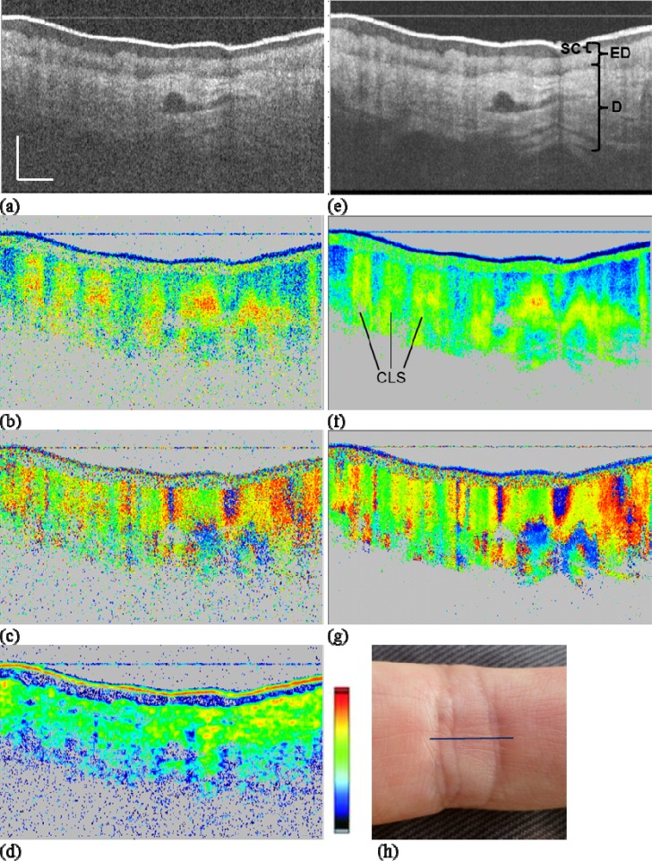
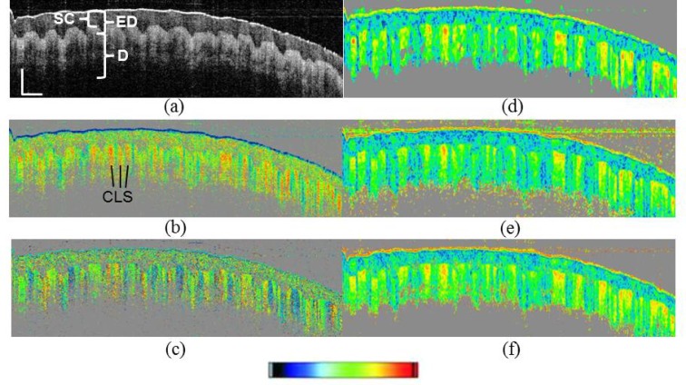
We recently reported on a new swept source polarization sensitive optical coherence tomography system and its application to skin imaging [1]. In three of the tomographic images of that paper (Figs. 4, 5, and 6) two skin layers were wrongly labeled: epidermis (E) and dermis (D) were interchanged. We replace these figures by the correctly labeled figures, Fig. 1 , Fig. 2 , and Fig. 3 , respectively. The text sections of the paper are not affected by that error.
Fig. 1.
(Replacement for Fig. 4 in original manuscript [1]) PS-OCT images of human skin. Proximal interphalangeal joint of middle finger (PIP) region. (a)–(d) single frame images; (e)–(g) average of 15 frames. (a), (e) reflectivity (log scale); (b), (f) retardation (color scale: 0–90°); (c), (g) axis orientation (color scale: −90–+90°); (d) DOPU (color scale: 0–1), 2D DOPU window (12(x) x 6(z) pixels or 55 x 38 µm2); (h) photo of imaged area, line shows approximate B-scan position. Scale bar dimensions: 0.5 mm (x: geometrical distance; z: optical distance). SC, stratum corneum; ED, epidermis; D, dermis; CLS, “column” like structure.
Fig. 2.
(Replacement for Fig. 5 in original manuscript [1]) PS-OCT images of human skin. Nail fold region. (a) reflectivity (log scale); (b) retardation (color scale: 0–90°); (c) axis orientation (color scale: −90–+90°); (d)–(f) DOPU (color scale: 0–1). (d) 2D DOPU window (16(x) x 7(z) pixels or 96 x 44 µm2); (e) 3D DOPU window (8(x) x 4(y) x 3(z) pixels or 48 x 48 x 19 µm3); (f) 3D DOPU window (5(x) x 5(y) x 3(z) pixels or 30 x 60 x 19 µm3). Scale bar dimensions: 0.5 mm (x: geometrical distance; z: optical distance). SC, stratum corneum;, ED, epidermis; D, dermis.
Fig. 3.
(Replacement for Fig. 6 in original manuscript [1]) PS-OCT images of human skin. Fingertip region. (a) reflectivity (log scale); (b) retardation (color scale: 0–90°); (c) axis orientation (color scale: −90–+90°); (d)–(f) DOPU (color scale: 0–1). (d) 2D DOPU window (6(x) x 12(z) pixels or 48 x 75 µm2); (e) 3D DOPU window (4(x) x 4(y) x 6(z) pixels or 32 x 32 x 38 µm3); (f) 3D DOPU window (2(x) x 6(y) x 6(z) pixels or 16 x 48 x 38 µm3). Scale bar dimensions: 0.5 mm (x: geometrical distance; z: optical distance). SC, stratum corneum;, ED, epidermis; D, dermis; CLS, “column” like structure.
References and links
- 1.Bonesi M., Sattmann H., Torzicky T., Zotter S., Baumann B., Pircher M., Götzinger E., Eigenwillig C., Wieser W., Huber R., Hitzenberger C. K., “High-speed polarization sensitive optical coherence tomography scan engine based on Fourier domain mode locked laser,” Biomed. Opt. Express 3(11), 2987–3000 (2012). 10.1364/BOE.3.002987 [DOI] [PMC free article] [PubMed] [Google Scholar]


