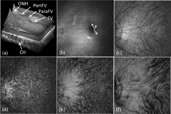
Fig. 7.
In vivo large field of view retina and choroid imaging results after compensation. (a) 3D volumetric image; (b) retinal fundus image obtained though integrating between 20 and 250 mm above the RPE layer; (c) choroidal fundus image obtained through integrating between 10 and 250 mm below the RPE layer; (d-f) depth resolved choroid fundus image obtained through integrating from 5 to 30 μm, 30 to 100 μm and 100 to 250 μm below the RPE layer, respectively. See also Media 1 (24.7MB, AVI) .

