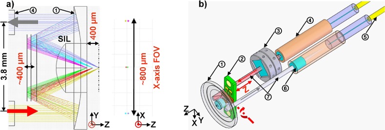
Fig. 1.
Endomicroscope schematic. (a) Cross-sectional view of dual axes architecture shows ray-trace simulation in ZEMAX for achieving 800 μm (width) × 400 μm (depth) images. (b) Optical circuit design for vertical cross-sectional imaging with fiber-coupled achromatic collimators. Components: (1) aluminum coated parabolic mirror with solid immersion lens (SIL) in center; (2) MEMS mirror with PCB holder; (3) prism with holder; (4) achromatic doublet lens based collimator; (5) single mode fiber; (6) achromatic lens; (7) illumination (red) and collection beam (gray).

