Fig. 5.
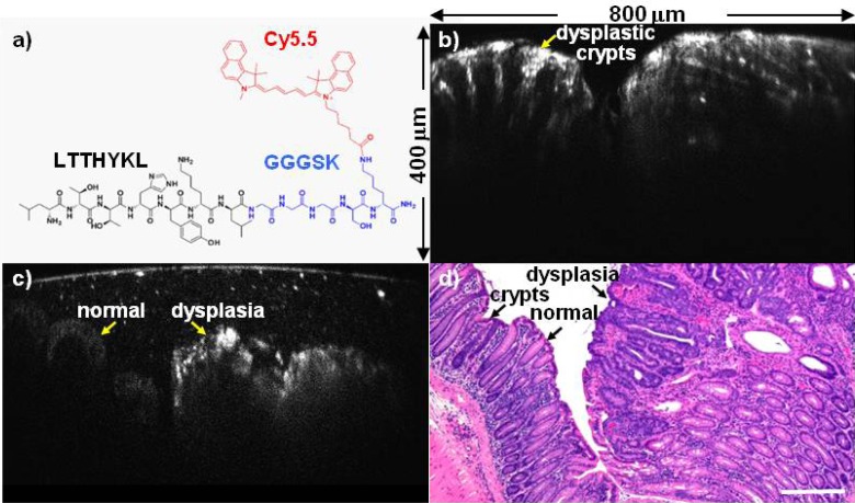
Vertical cross-sectional image of colonic dysplasia. (a) Chemical structure of LTTHYKL peptide (black) with GGGSK linker (blue) and Cy5.5 fluorophore (red). (b) NIR fluorescence image from CPC;Apc mouse colon ex vivo shows vertically oriented dysplastic crypts. (800 × 400 μm2 FOV) (c) The border between normal colonic mucosa and dysplasia shows increased contrast from specific binding of the LTT*-Cy5.5 peptide. (800 × 400 μm2 FOV) (d) Corresponding histology (H&E), scale bar 200 µm.

