
Figure 2.
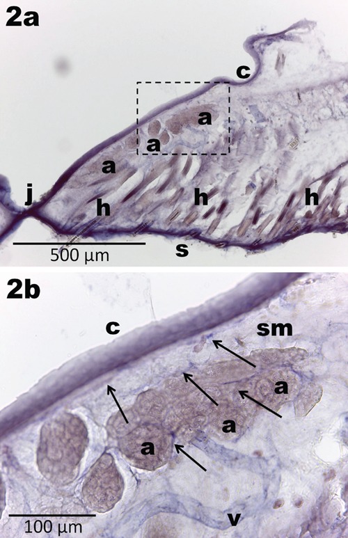
Parasagittal section through the rat upper eyelid on 14th postnatal day, RA group, NADPH-diaphorase staining. a) Shrunken acini of Meibomian glands (a) lie near the conjunctival surface (c) of the eyelid; skin (s); hairs (h); junction between upper and lower eyelid (j); blood vessel (v); scale bar: 500 µm. b) Inset from a: blood vessels accompanied by nerve fibers (arrows) are running mostly in submucosa (sm); and rarely between acini of the Meibomian glands (a); scale bar: 100 µm.