
Figure 2.
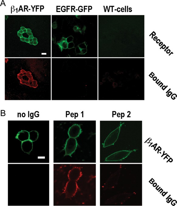
Specificity of IgG-β1ARs colocalization at specificity cut-off. (A) β1AR- (top) and IgG-associated fluorescence (bottom) in wild-type cells (right) and cells expressing YFP-fused human β1ARs (left) or GFP-fused human EGF receptors (middle), all stained with patient IgG at 1:500 dilution (26.8 mg/L). Images obtained at 400-fold magnification, size bar of 30 μm at top left applies to all panels. (B) β1AR- (top) and IgG-associated fluorescence (bottom) in cells expressing YFP-fused human β1ARs and stained with secondary antibody alone (left) or human autoimmune IgG (26.8 mg/L) pre-absorbed with a 1000-fold molar excess of peptide analogues of the first (middle) or the second (right) extracellular loop of the human β1AR. Images obtained at 630-fold magnification. Size bar of 30 μm at top left applies to all panels.