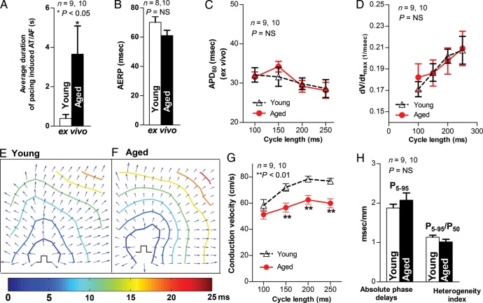
Figure 4.
Increased pacing-induced AT/AF and slowed CV in aged rabbit LA ex vivo. (A–C) Pooled data of inducibility and duration of pacing-induced AT/AF, AERP, and APD60 in Langendorff-perfused aged and young rabbit LA (*P < 0.05, P = NS vs. young, respectively). (D) Summarized data for unchanged CL-dependent dV/dtmax in aged rabbit LA (P = NS). (E–F) Representative isochronal maps from young and aged rabbit hearts subjected to pacing at a CL of 200 ms (π indicates the pacing sites). (G) Summarized optical mapping CV data show that aged rabbits exhibited a CL-dependent, slower conduction (**P < 0.01 vs. young controls). (H) Analysed optical mapping data of unchanged absolute phase delays (conduction inhomogeneity, P5–95) and heterogeneity index (P5–95/P50) between young and aged LA (P = NS).

