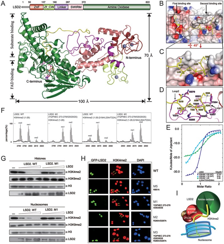
Figure 1.
Structural insight into the substrate recognition of LSD2. (A) Overall structure of LSD2-H3K4M(1-26) is shown as a ribbon representation in two different views. The H3K4M peptide is colored in yellow. FAD is shown in stick representation (purple) and three zinc atoms are shown as grey balls. Schematic representation of the domain structure of human LSD2 with boundaries for each domain is indicated above the structure. The same color scheme is used in all structure figures of LSD2. (B) LSD2 is shown in electrostatic potential surface representation and the H3K4M peptide is shown in ribbon representation, with two substrate-binding sites highlighted by rectangles. (C) A zoom-in view of the interaction between the H3K4M peptide and the second binding site. (D) A close-up view as shown in C. Residues of H3K4M and LSD2 are labeled and colored in yellow and purple, respectively. Hydrogen bonds are shown as dashed lines. Two critical loops for the second binding site formation are indicated. (E) Superimposed ITC enthalpy plots for the binding of histone H3K4M peptides (syringe) to purified wild-type LSD2 protein (cell) with the estimated binding affinity (Kd) listed. (F) In vitro histone demethylation assay using H3K4me2 peptides (residues 1-26, WT and Q19A/L20A/T22A) as substrates. LSD2 was used at a high protein concentration. MALDI-TOF mass spectrometry analyses of demethylation by LSD2 wild-type or mutant proteins are shown. (G) In vitro histone demethylation assay using histones and nucleosomes as substrates, respectively. LSD2 wild-type or mutant proteins were used in three different concentrations and the reactions were monitored by immunoblotting with indicated antibodies. (H) Immunofluorescence analysis of H3K4me2 demethylase activities in 293T cells transfected with GFP fusion proteins as indicated. Green, GFP fusion protein; red, H3K4me2; blue, DAPI staining of nuclei. Transfected cells are indicated by arrows. The reduced levels of H3K4me2 were observed in cells expressing the wild-type LSD2, but not its mutants. (I) The catalytic cavity in the AO domain is the first substrate-binding site, which binds to the N-terminus of histone H3K4me2 for demethylation. The linker region forms the second binding site away from the catalytic cavity. This additional interaction is important for histone H3 recognition and essential for demethylation activity of LSD2.

