
Fig. 2.
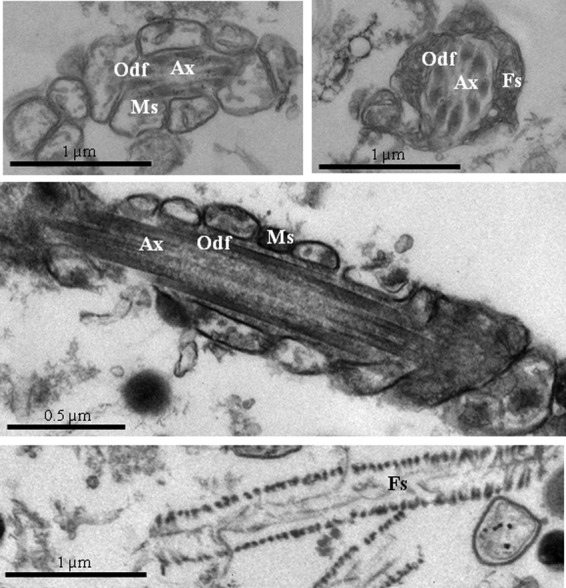
Electron microscope pictures of human sperm tail fractions isolated by means of sonication and sucrose gradient ultracentrifugation. All the typical tail structures were observed: axonemes (Ax), outer dense fibers (Odf), mitochondrial sheaths (Ms), and fibrous sheaths (Fs).