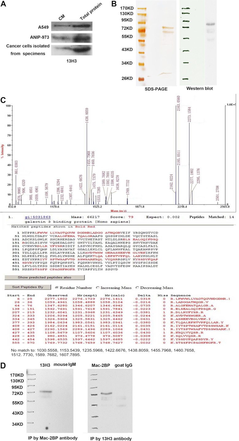
Fig. 2.
Mac-2BP is identified as the antigen of mAb 13H3. A, Western blot analysis (20 μg protein) of the conditioned medium from A549, ANIP-973 and lung cancer tissues using 13H3. B, Left, specific tumor antigen that was visualized on silver stained method; Right, validated by Western blotting using 13H3 as the primary antibody. C, Mass spectrometric analysis of the tumor antigen. Upper panel showed the peptide mass fingerprint of Mac-2BP; lower panel showed the result for database search showing the detected peptide fragments and the peptide coverage. D, The immunoprecipitate by anti-Mac-2BP could be recognized by 13H3.

