Fig. 4.
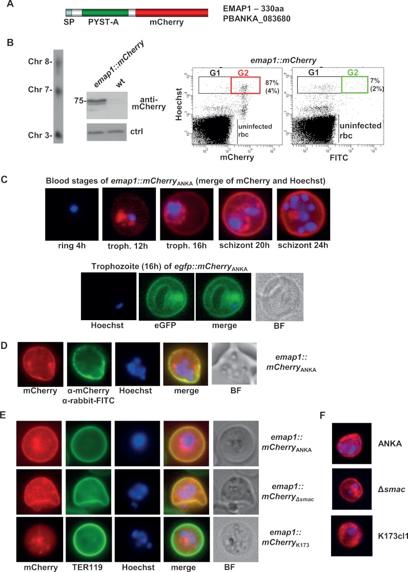
EMAP1 (PBANKA_083680) is associated with the irbc membrane of P. berghei ANKA irbcs. A, schematic of mCherry-tagged EMAP1 showing the location of the predicted signal peptide (sp) and the P. yoelii subtelomeric A domain (PYST- A). B, analysis of emap1::mCherry parasites. Left panel: Southern analysis of separated chromosomes shows integration of the tagging construct into chromosome 8. Middle panel: Western analysis of EMAP1::mCherry expression using anti-mCherry antibodies. As a loading control (ctrl) for wild-type (wt) parasites, we used the aspecific reaction of the antibodies with a ∼20 kDa parasite protein. Right panel: FACS analysis of mCherry-expressing schizonts. In the first dot plot, the irbcs are selected based on Hoechst and mCherry fluorescence. An average percentage of 87% (+ 4; n = 5; Gate 2) of the total number of schizonts (Gate 1) is mCherry positive. In the second dot plot, the irbcs were selected based on Hoechst and FITC fluorescence after staining with primary anti-mCherry antibodies and secondary FITC antibodies. An average percentage of 7% (+ 2; n = 2; Gate 2) of the total number of schizonts (Gate 1) is FITC positive. Gate 1 (G1): mature schizonts (8–16N); Gate 2: mCherry- or FITC-positive schizonts. C, irbc membrane location of EMAP1 in live blood stages of ANKAwt as shown by fluorescence microscopy of mCherry- (red) or eGFP (green)-tagged EMAP1. Nuclei are stained with Hoechst (blue). D, detection of EMAP1::mCherry at the surface of live irbcs via staining with primary anti-mCherry antibodies and secondary FITC antibodies (green). BF, bright field. E, irbc membrane location of EMAP1 in live blood stages of ANKAwt and ANKAΔsmac as shown by fluorescence microscopy of mCherry-tagged EMAP1 (red). In K173cl1 blood stages, EMAP1::mCherry shows a more diffuse and patchy localization in the cytoplasm of irbcs, with no distinct irbc membrane localization. The irbc surface membrane is stained with TER119 antibody (green), and nuclei are stained with Hoechst (blue). F, localization of EMAP1 in fixed irbcs as determined via immunofluorescence microscopy after staining with primary anti-EMAP1 antibodies and secondary Alexa 594 antibodies. These analyses confirm the association of EMAP1 with the irbc membrane in ANKAwt and ANKAΔsmac parasites and the more diffuse or patchy localization in K173cl1.

