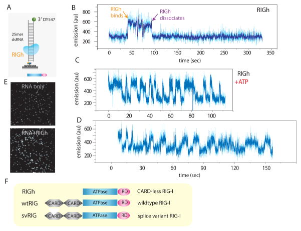
Fig. 1. PIFE visualization of RIG-I binding and translocation.
A. dsRNA (25mer) with single fluorophore (DY547) was tethered to surface via biotin-neutravidin. B. Addition of RIGh (CARD-less mutant) resulted in an abrupt increase in emission of the fluorophore, indicating RIGh binding due to PIFE (protein induced fluorescence enhancement). C, D. Addition of RIGh with ATP induced periodic fluctuation of fluorophore. E. The effect of PIFE is visible on the single molecule imaging surface i.e. fluorescence become substantially brighter upon adding RIGh protein. F. Schematic representation of three RIG-I variants used in this study; wtRIG (RIG-I wild type), RIGh (CARD-less RIG-I) and svRIG (RIG-I splice variant) is shown.

