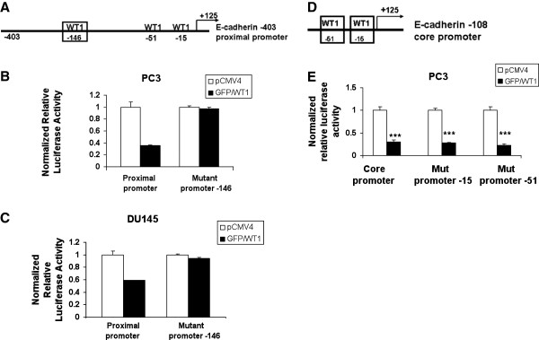
Figure 4.

WT1 binding site located at −146 bp is required for E-cadherin promoter repression. (A) Schematic diagram of the E-cadherin proximal promoter showing the −146 bp WT1 binding site (box). (B) PC3 and (C) DU145 cells were transiently cotransfected with GFP/WT1 and either with wild type or mutant proximal promoter containing a mutated −146 WT1 binding site. (D) Schematic diagram of the E-cadherin core promoter showing −15 and −51 bp WT1 binding sites (boxes). (E) PC3 cells were transiently cotransfected with GFP/WT1 and either with wild type or mutant core promoters containing a mutated −15 or −51 WT1 binding site. Luciferase activity was measured and normalized as described in Figure 3. Experiments were repeated three times in triplicate. Data are reported relative to luciferase activity of pCMV4. Significance was determined by student’s t-test comparing GFP/WT1 transfected cells relative to pCMV4 transfected cells (***p ≤ 0.001) in three independent experiments.
