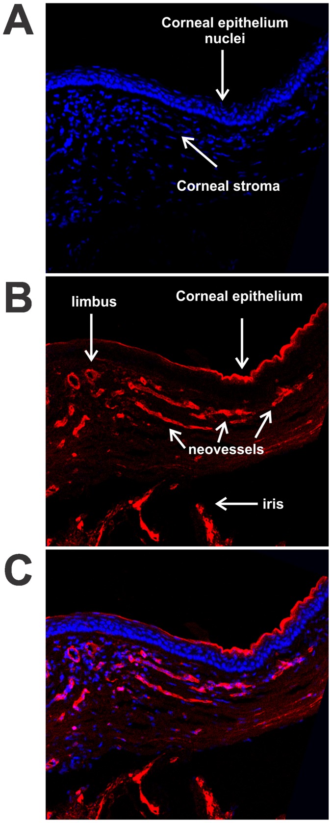
Figure 5. Localization of corneal neovessels with isolectin IB4 .
. Cryosections of temporal corneas were prepared 14 days after implantation with a 20% 7KCh wafer. The sections were labeled with AlexaFluor 568 Isolectin IB4 (red) and the nuclei stained with DAPI (blue). A. DAPI stained section demonstrating the location of the corneal stroma and epithelium. B. Isolectin IB4 labeling of neovessels, corneal epithelium and iris. C. Combined image. Arrows mark the different structures.

