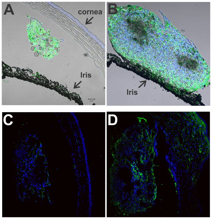
Figure 7. Localization of CD68 positive cells and VEGF in Ch and 7KCh implants.
Rat eyes were implanted with 20% Ch and 7KCh wafers and cryosections prepared 14 days post implantation. The sections were labeled with AlexaFluor 488 (green) for anti-CD68 or anti-VEGF and the nuclei stained with DAPI (blue). A. Ch implant, anti-CD68. B. 7KCh implant, anti-CD68. C. Ch implant, anti-VEGF. D. 7KCh implant, anti-VEGF.

