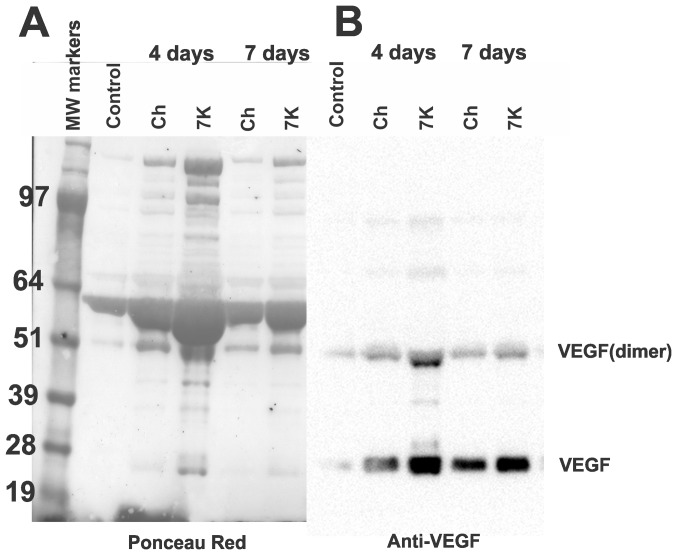
Figure 8. SDS-PAGE and immunoblotting of AH from Ch and 7KCh implants.
AH (5 µl) from control (un-implanted eye), Ch and 7KCh implanted eyes were separated by SDS-PAGE and blotted. A. Ponceau S red stained blot demonstrating the difference in protein content between samples. B. Anti-VEGF labeling of blot.

