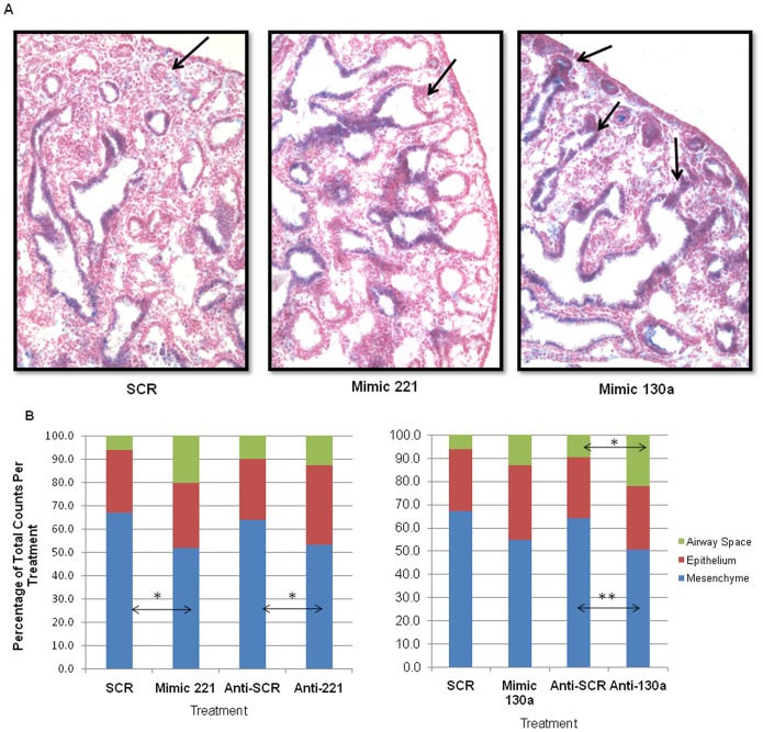
Figure 4. Epithelial and morphological changes observed in lungs with anti-miR and mimic treatments.
(A) Mimic 130a alters epithelial expression of Sox2, a transcription factor expressed in proximal but not distal airways. Immunohistochemistry was done on sections of cultured lung using Sox2 antibody. Mimic 130a lungs had nuclear Sox2 expression in distal airway epithelium (arrows), which was minimal or absent in SCR and Mimic 221 lungs. Images were taken at 20× magnification. The figure shows representative images from N = 3 lungs per condition. (B) Morphometric analysis showed altered lung morphology in mimic and anti-miR-treated lungs. Point count analysis was done by placing a 50μm grid on histological sections. Mimic 221 had a 15.2% decrease and anti-221 a 10.5% decrease in mesenchymal area compared to controls. Mimic 130a treatment showed a trend towards decreased mesenchymal area and an increase in epithelial and airway space area. Anti-130a treatments caused a 13.4% decrease in mesenchymal area and a 12.4% increase in airway space area. Mean ± SEM of N ≥3, * p<0.05, **p<0.01. N≥40 tissue sections each separated by at least 18µm from ≥3 lungs per condition.

