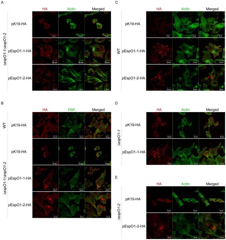
Figure 2. Localization of EspO1-1 and EspO1-2 in EHEC-infected cells.
(A) HeLa cells were infected with ΔespO1-1ΔespO1-2 carrying pK19-HA or ΔespO1-1ΔespO1-2 carrying a plasmid with the gene for either HA-tagged EspO1-1 (pEspO1-1-HA) or EspO1-2 (pEspO1-2-HA) and, at 4 h post-infection, were analyzed by immunofluorescence staining. Cells infected with ΔespO1-1ΔespO1-2 carrying pEspO1-1-HA or ΔespO1-1ΔespO1-2 carrying pEspO1-2-HA were processed for confocal laser scanning microscopy using Alexa 488-conjugated phalloidin to visualize actin filaments (green), and anti-HA antibody to visualize HA-tagged proteins (red). (B) HeLa cells were infected with WT carrying pK19-HA, or ΔespO1-1ΔespO1-2 carrying pK19-HA, pEspO1-1-HA or pEspO1-2-HA. Epithelial cells were infected with these strains for 4 h and immunofluorescence-stained with anti-FAK antibody to visualize focal adhesions (green) and anti-HA antibody (red). (C) HeLa cells were infected with WT carrying pK19-HA, pEspO1-1-HA or pEspO1-2-HA. (D) HeLa cells were infected with ΔespO1-1 carrying pK19-HA or pEspO1-1-HA. (E) HeLa cells were infected with ΔespO1-2 carrying pK19-HA or pEspO1-2-HA. Epithelial cells were infected with these strains for 4 h and immunofluorescence-stained with Alexa 488-conjugated phalloidin (green) and anti-HA antibody (red).

