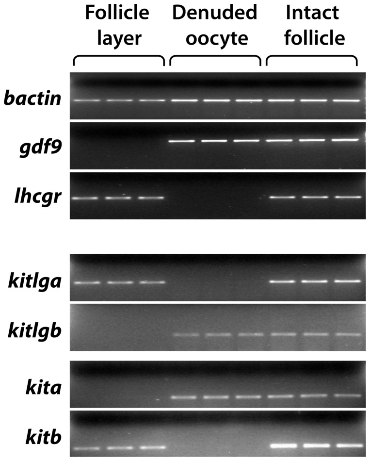
Figure 1. Distribution of the Kit system within the zebrafish follicle.
The somatic follicle layer was separated from the oocyte followed by RNA extraction and semi-quantitative PCR detection in the two compartments (follicle layer and denuded oocyte). Each sample represents the total RNA pooled from 5 follicles. The housekeeping gene bactin was used as the internal control for all samples, whereas gdf9 and lhcgr were used as the markers for denuded oocytes and somatic follicle layers, respectively.

