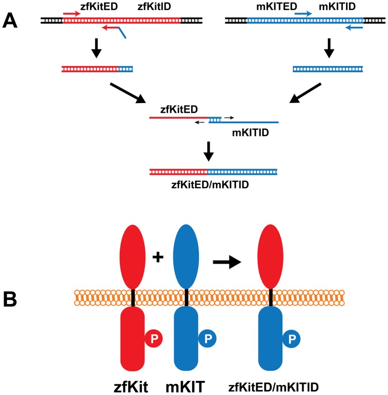
Figure 6. Schematic illustration of plasmid construction for expressing chimeric receptors.
The mouse Kit receptor is shown in blue and zebrafish Kita or Kitb is in red. The extracellular domain of zebrafish Kita or Kitb (zfKitED) and intracellular domain of mouse KIT (mKITID) were amplified by PCR followed by extension of the PCR products on each other to generate a DNA fragment (A) coding for the fusion protein zfKitED/mKITID (B). “P” represents the phosphorylated tyrosine site in the intracellular domain that is recognized by the antibody.

