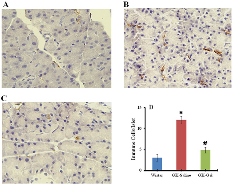
Figure 4. Immunohistochemical staining of macrophage infiltration marker (CD68) in pancreatic islets (IHC ×400) of: (A) Wister rat (B) GK-Saline group showing abundant CD68 labeling representing rich macrophage infiltration (C) GK-Gel group showing normal organization of islets with minimal macrophage infiltration.
(D) Quantification of immune cells/pancreatic islets by immunohistochemistry. *, P<0.05; compared to wistar rats groups. #, P<0.05; compared to GK-Saline group. n = 3 for wistar rats, n = 4 for both groups of GK rats.

