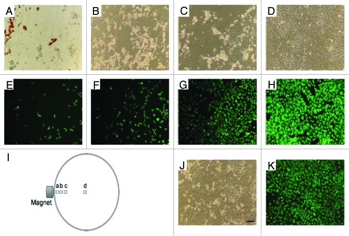
Figure 2. Phase contrast (A–D) and fluorescence (stained with Calcein AM) microscope images of HeLa cells incubated for 24 h with doxorubicin-loaded magnetic LiPSi, showing magnetically guided (H) and without guidance (J and K) delivery of doxorubicin. The position of each image relative to the external magnet is depicted in I). Scale bar for all images is 100 µm. Reprinted with permission from reference 48.

An official website of the United States government
Here's how you know
Official websites use .gov
A
.gov website belongs to an official
government organization in the United States.
Secure .gov websites use HTTPS
A lock (
) or https:// means you've safely
connected to the .gov website. Share sensitive
information only on official, secure websites.
