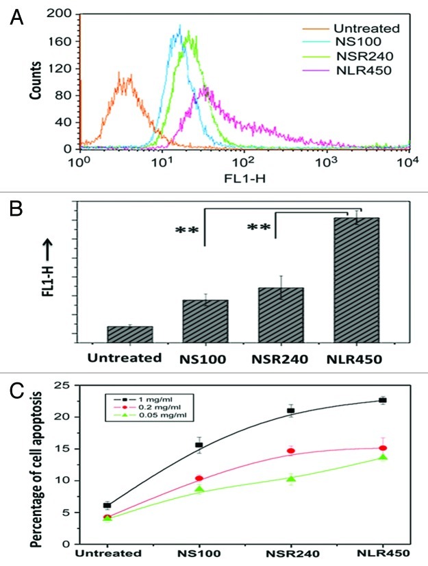
Figure 4. (A) Quantification of cellular uptake and apoptosis of different shaped PSiO2 by flow cytometry analysis of different prepared PSiO2 nanoparticles: sphere-shaped, NS (100 nm), short rod-shaped, NSR (240 nm) and long rod-shaped, NLR (450 nm). (B) Quantitative measurement (fluorescence) of particle numbers in cells (data are presented as mean ± s.d; statistical significance for the comparison of the number of internalized particles between different shaped particles, **p < 0.1). (C) Influence of different concentrations of shaped nanoparticles on early apoptosis of A375 cells. Reprinted with permission from reference 77.

An official website of the United States government
Here's how you know
Official websites use .gov
A
.gov website belongs to an official
government organization in the United States.
Secure .gov websites use HTTPS
A lock (
) or https:// means you've safely
connected to the .gov website. Share sensitive
information only on official, secure websites.
