
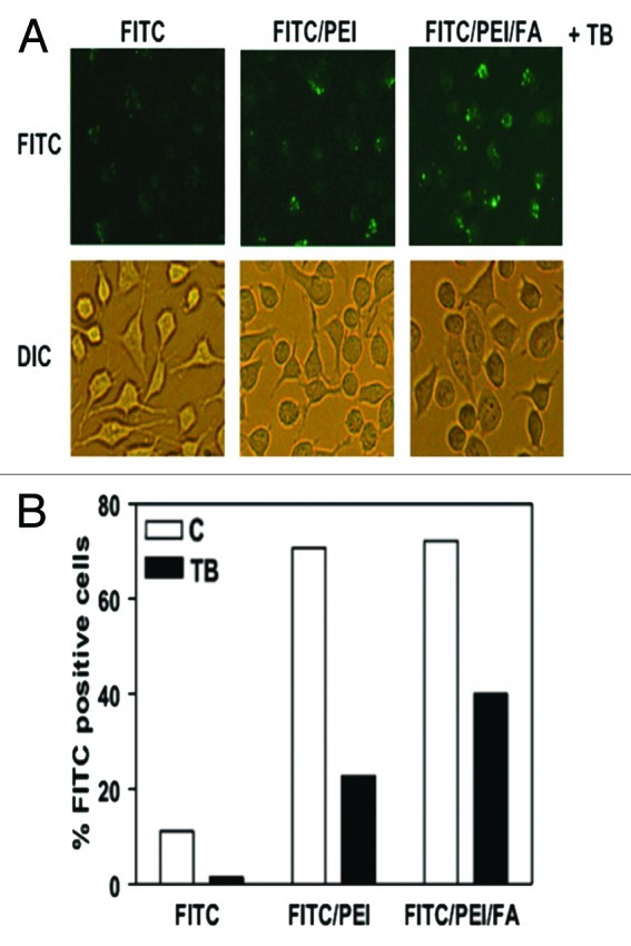
Figure 6. Conjugation of FA with PSiO2 nanoparticle showed improved endocytosis in HeLa cells. HeLa cells treated with PSiO2 nanoparticles at 10 µg/ml for 2 h and the extracellular binding of the nanoparticles was quenched with trypan blue (FITC, pristine FITC-labeled particle; FITC/PEI, PEI-functionalized FITC-labeled particle; FITC/PEI/FA, FA-conjugated PEI-functionalized FITC-labeled particle). Fluorescence microscopy was used to image FITC-labeled particles inside the cells and differential interference contrast microscopy for cellular morphology evaluations (A). Flow cytometry was used to detect the number of HeLa cells with endocytosed PSiO2 nanoparticles in control or trypan blue quenched cells (B). Reprinted with permission from reference 53.