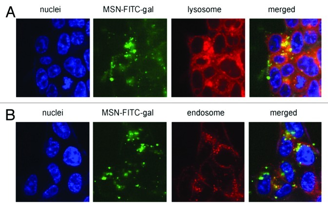
Figure 7. Confocal microscopy images after 6 h incubation of living HCT-116 colorectal cancer cells with FITC-galactose-modified PSiO2 nanoparticles ( = MSN-FITC-gal) at 37°C. Merged pictures of both section A and B indicated the co-localization (yellow) of FITC-nanoparticles (green) with lysosomal or endosomal markers, respectively. Reprinted with permission from reference 113.

An official website of the United States government
Here's how you know
Official websites use .gov
A
.gov website belongs to an official
government organization in the United States.
Secure .gov websites use HTTPS
A lock (
) or https:// means you've safely
connected to the .gov website. Share sensitive
information only on official, secure websites.
