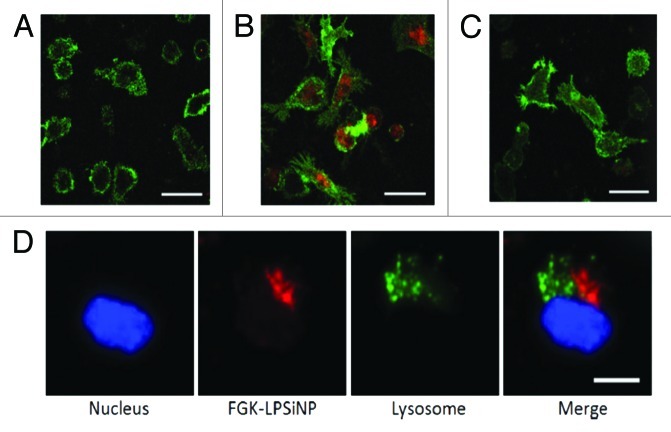
Figure 8. Fluorescence microscope images of mouse bone marrow derived dendritic cells treated with (A) LPSiNPs or (B) FGK-LPSiNPs for 1.5 h at 37°C. (C) 30 min pretreatment of the bone marrow-derived dendritic cells (green) with free FGK45 inhibits uptake of FGK-LPSiNPs incubated with the cells for 1.5 h at 37°C. FGK-LPSiNPs were detected by their intrinsic visible/near-infrared photoluminescence (red, λex = 405 nm and λem = 700 nm). The scale bars are 40 µm. (D) Distribution of FGK-LPSiNPs bone marrow-derived dendritic cells after 1.5 h incubation at 37°C. The lysosomes are stained in green (LysoTracker; Invitrogen), the nucleus in blue and FGK-LPSiNPs in red. The scale bar is 10 µm. Reprinted and modified with permission from reference 116.

An official website of the United States government
Here's how you know
Official websites use .gov
A
.gov website belongs to an official
government organization in the United States.
Secure .gov websites use HTTPS
A lock (
) or https:// means you've safely
connected to the .gov website. Share sensitive
information only on official, secure websites.
