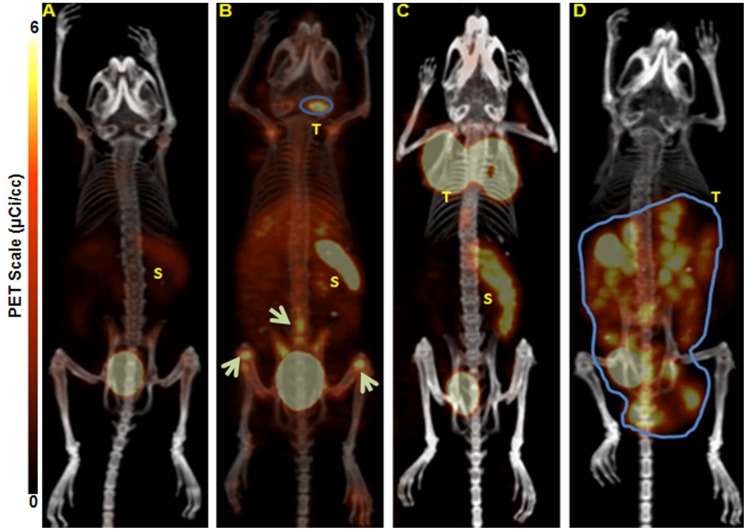
Figure 4. Representative maximum intensity projection (MIP) small animal PET/CT images.
A. non-tumor KaLwRij control mouse. B. a small sized, non-palpable, early stage subcutaneous (s.c.) 5TGM1 murine tumor in the nape of the neck inoculated without the use of matrigel (tumor SUV 2.24). White arrows point to suspected tumor cells and associated tumor supporting cells in the BM of the long bones and spine. C. matrigel assisted s.c. 5TGM1 tumor in the nape of the neck (tumor SUV 6.2). D. mouse injected intraperitoneally (i.p.) with 5TGM1 murine myeloma cells. All the mice were injected with 64Cu-CB-TE1A1P-LLP2A (0.9 MBq, 0.05 µg, 27 pmol) and were imaged by small animal PET/CT at 2 h post-injection. *All tumor bearing animals were SPEP (Serum Protein Electrophoresis) positive. T = Tumor; S = Spleen. N = 4/group.

