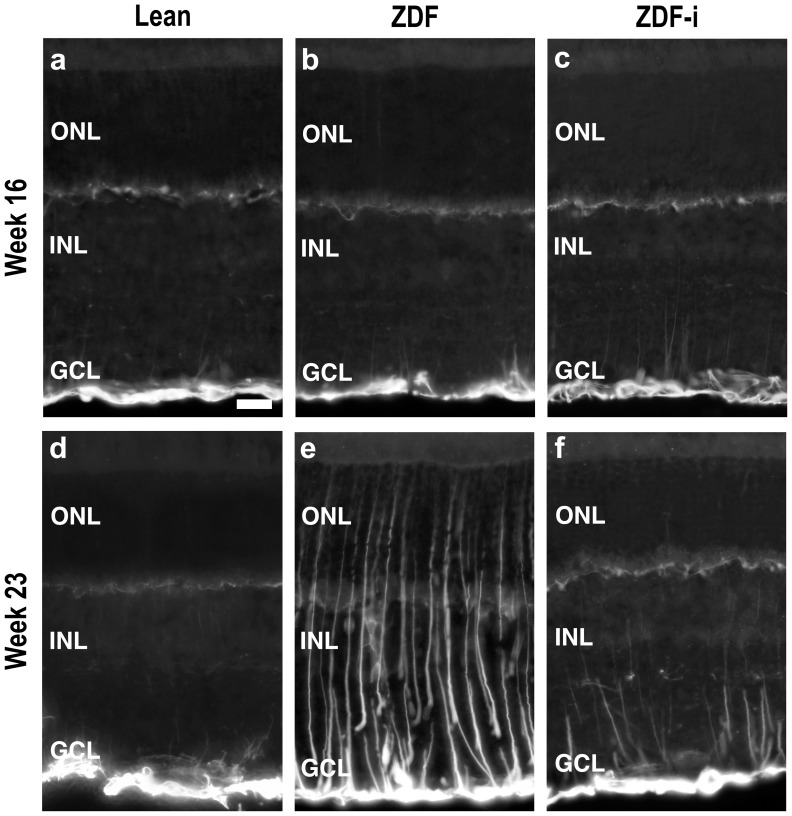
Figure 8. GFAP immunofluorescence.
Localization of GFAP in cryostat sections obtained from Lean (a), ZDF (b) and insulin treated ZDF (c) rats at 16 weeks of age; and from Lean (d), ZDF (e) and insulin treated ZDF (f) rats at 23 weeks of age. ZDF-i, insulin-treated ZDFs; ONL, outer nuclear layer; INL, inner nuclear layer; GCL, ganglion cell layer. Scale bar = 20 µm.

