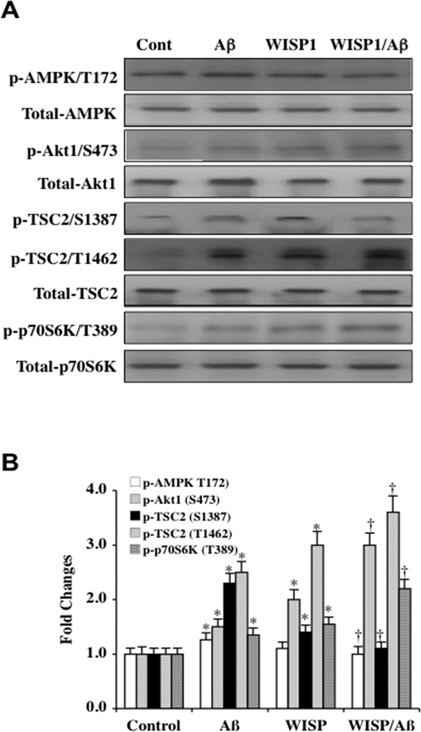
Fig. (1). WISP1 modulates post-translational phosphorylation of AMPK, Akt1, TSC2, and p70S6K during Aβ exposure.
(A) Equal amounts of microglial protein extracts (50 µg/lane) were immunoblotted at 6 hours following Aβ exposure with total and anti–phospho p-AMPK (Thr172), p-Akt1 (Ser2448), p-TSC2 (Ser1387/Thr1462), and p-p70S6K (Thr389) antibodies. WISP1 (10 ng/ml) was applied to microglia cultures 1 hour prior to Aβ exposure (10 µM). During Aβ exposure, phosphorylation of AMPK, TSC2 Ser1387, TSC2 Thr1462 were increased. In contrast, WISP1 decreased phosphorylation of AMPK and TSC2 Ser1387 in the presence of Aβ. WISP1 increased phosphorylation of Akt1, TSC2 Thr1462, and p70S6K during Aβ exposure. Representative images for western blot analysis are illustrated. (B) Quantitative analysis of western analysis from 3 experiments was performed using the public domain NIH Image program (US National Institutes of Health at http://rsb.info.nih.gov/nih-image/) (*P < 0.01 vs. Control; †P<0.01 vs. Aβ treated alone). Control = untreated microglia. Each data point represents the mean and SD.

