Fig. 4.
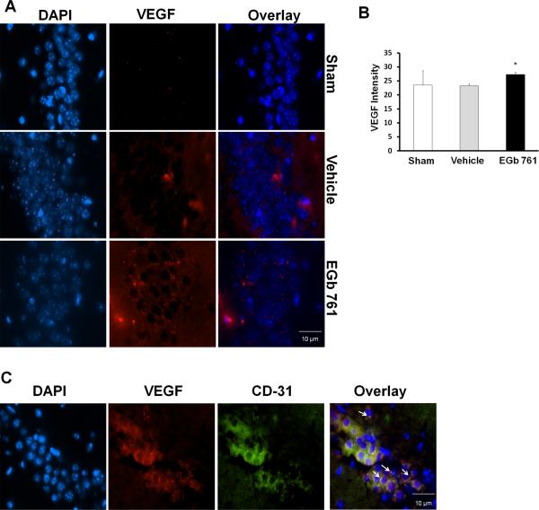
EGb 761 upregulates VEGF expression in the CA-1 region of the hippocampus. Hippocampal sections (paraffin) of mice pretreated with EGb 761 for 7 days and subjected to 8-minutes of global ischemia were used in this immunofluorescence assay. (A) VEGF expression was visualized by immunofluorescent staining with a specific rabbit polyclonal antibody followed by secondary anti rabbit IgG (red), and DNA was stained blue (DAPI). (B) VEGF expression was increased in EGb 761 pretreated mice as compared to vehicle group. (C) Double staining for VEGF localization was visualized by a specific anti rabbit polyclonal antibody followed by secondary anti rabbit IgG (red) and CD-31 mouse monoclonal antibody for endothelial cell marker followed by secondary anti-rat IgG (green). The arrows in the figure show overlapping of CD-31 (green) on HO1 (red), thereby giving out yellow fluorescence. VEGF was observed to be expressed in endothelial cells. Magnified view, 60X; * vs. vehicle; p < 0.05.

