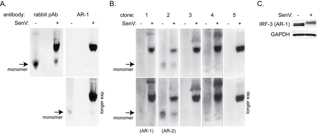
Figure 4.
A. Native PAGE followed by immunoblot analysis for IRF-3 in SupT1 cells infected with SenV for 18 hrs. A rabbit polyclonal antibody or AR-1 mAb was used to detect IRF-3. Arrows indicate monomeric IRF-3. Short (top) and long (bottom) film exposures are shown.
B. Native PAGE followed by immunoblot analysis for IRF-3 using supernatants from five hybridoma clones. Clone number 1 represents AR-1 mAb, while clone number 2 represents AR-2 mAb. Short and long film exposures of each immunoblot assay are shown as in 4A.
C. SDS-PAGE followed by immunoblot analysis for IRF-3 (AR-1) for loading control.

