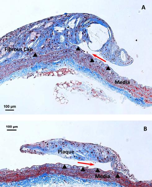
Figure 3. Histological demonstration of delamination surface location.
A. Masson's Trichrome staining of a longitudinal section of a partially delaminated plaque from an apoE KO mouse (200×, scale bar=100 μm), leaving part of it attached to the underlying IEL (arrowheads). The crack front (red arrow) is located between the atherosclerotic plaque cap and its underlying IEL; B. A partially delaminated plaque from an apoE MMP-12 DKO mouse (200×, scale bar=100 Pm). Plaque separation occurred at the same interface. Collagen is stained blue, nuclei purple-black, and smooth muscle cells red.

