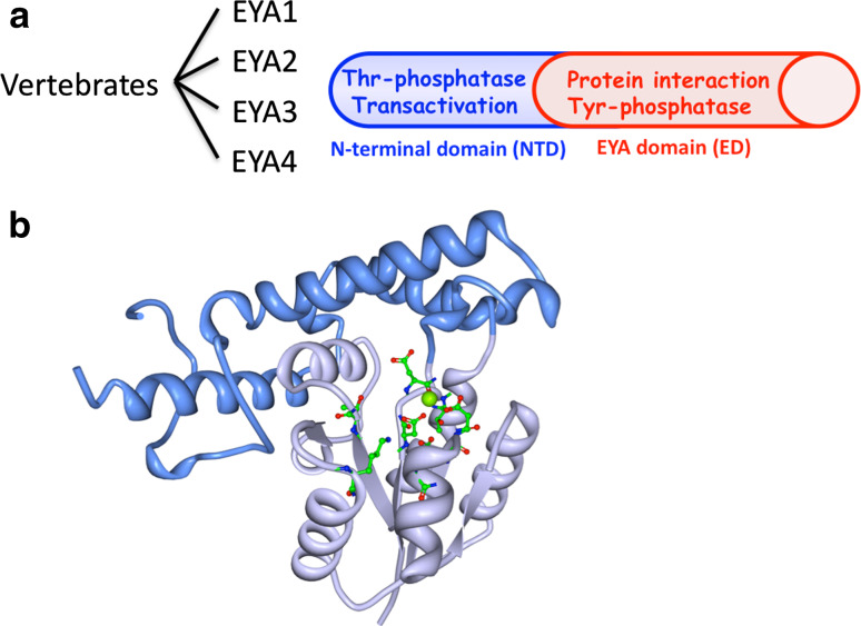
Fig. 1.
Molecular architecture of the EYA proteins. a There are four vertebrate EYA proteins. They have a well-conserved C-terminal domain called the EYA domain (ED) and a poorly conserved N-terminal domain (NTD). The ED participates in protein interactions, notably with the SIX family of proteins that are part of the PSEDN. The ED also has tyrosine phosphatase activity. The NTD has transactivation and threonine phosphatase activities. b The three-dimensional structure of the EYA2 ED as determined by X-ray crystallography [14]. It has two subdomains referred to as the core (light blue) and cap (dark blue) domains. The active site residues are shown in green. A divalent metal ion (green sphere) in the active site participates in the catalytic reaction

