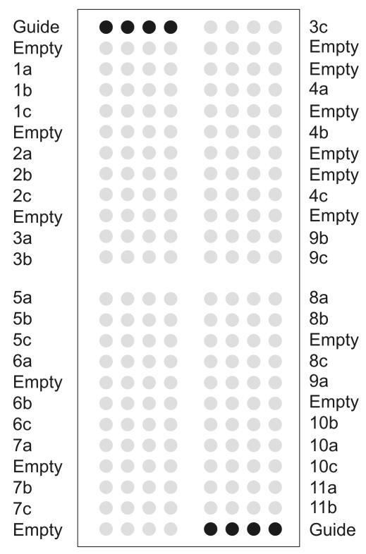
FIG. 1.
DNA chip layout. The spotted solid-phase PCR primer array contains 384 features, each of which is 190 to 200 μm in diameter with a spot-to-spot distance of 100 μm. The spotted solid-phase primers are represented as gray circles; their names are indicated on the left and right. The corresponding sequences are shown in Table 2. Black circles in the upper left and lower right corners are guide dots used for orientation and grid alignment during analysis. Dots with buffer only are referred to as empty. For reasons of clarity, each solid-phase primer is schematically indicated by three replicate dots; the real DNA chip layout contains 11 replicate dots for each primer.

