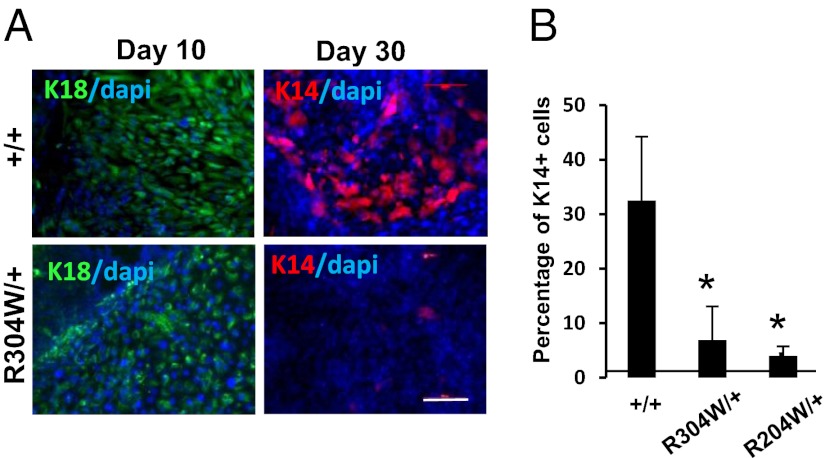
Fig. 1.
Impaired epidermal differentiation of EEC-iPSC. iPSC+/+, iPSCR204W/+, and iPSCR304W/+ cells were subjected to epidermal differentiation protocol in presence of BMP-4 and SB431542, and immunofluorescence for K18 and K14 after 15 d of differentiation (A) or analyzed by flow cytometry analysis for K18 and K14 after 25 d of differentiation (B). The data are an average of the percentage of K14+ cells ± SE from two independent iPSC clones. *P < 0.001. (Scale bar: 20 μm.)

