Fig. 2.
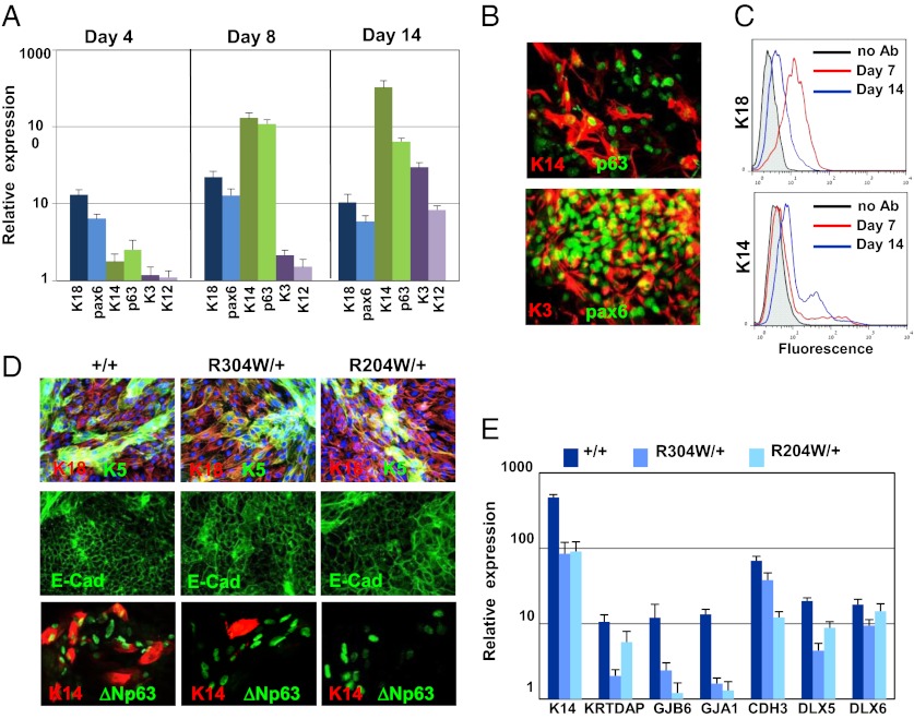
Impaired corneal epithelial commitment of iPSC lines. iPSC+/+ were seeded on collagen IV-coated dishes in corneal fibroblast-conditioned medium that was supplemented with BMP-4 for the first 3 d of differentiation. Cells were harvested at the indicated time points and subjected to real-time PCR analysis of ectodermal markers (pax6 and K18), corneal epithelial progenitor markers (K14 and p63), and markers of terminally differentiated corneal-epithelial cells (K3 and K12) (A). Cells were collected at day 14 of differentiation and subjected to coimmunofluorescent staining of p63 and K14 or pax6 and K3 (B). Flow cytometry analysis of iPSC+/+ that were harvested at days 7 and 14 of differentiation and stained with K18 or K14 antibodies is shown in C. iPSC+/+ and iPSCEEC lines were differentiated into corneal epithelial fate for 10 d (E). Immunostaining followed by fluorescent microscopy was performed for determining the expression of the indicated proteins (D), and real-time PCR analysis was performed for determining the relative expression of the indicated epithelial transcripts (E). (Magnification: B, 40×; D, 20×.)

