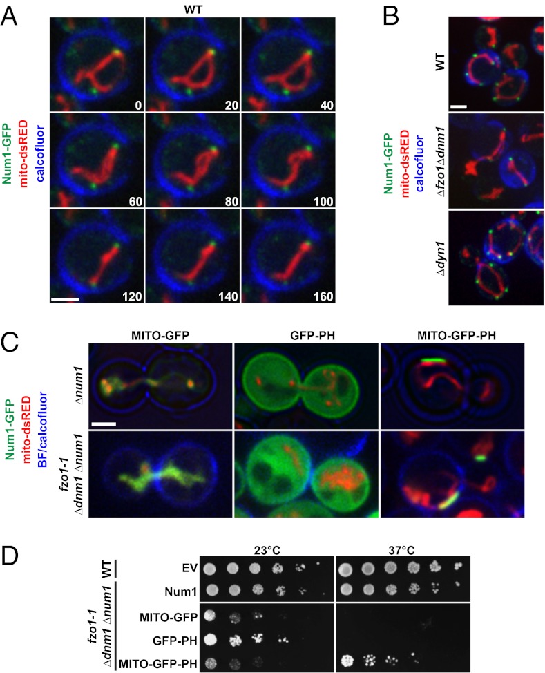
Fig. 2.
Num1 physically tethers mitochondria to the cell cortex. (A) Time-lapse images of calcofluor-stained wild-type yeast cells expressing Num1-GFP and mito-dsRED. A single focal plane is shown. Time is shown in seconds. (B) Num1-mediated tethering is independent of mitochondrial fusion and division and is independent of Num1’s role in dynein-mediated nuclear migration. Images of calcofluor-stained wild-type, Δdyn1, and Δfzo1 Δdnm1 cells expressing Num1-GFP and mito-dsRED are shown. (C) Images of Δnum1 cells (Upper) and fzo1-1 Δdnm1 Δnum1 cells (Lower) expressing MITO-GFP, GFP-PH, or MITO-GFP-PH as indicated. Δnum1 cells were grown at 30 °C before imaging, and fzo1-1 and fzo1-1 Δnum1 cells were imaged after the cells were shifted to the nonpermissive temperature (37 °C) for 5 h. The cell cortex was visualized using bright-field (BF) imaging (Upper) or by staining the cells with calcofluor before imaging (Lower). Single focal planes are shown. (D) Expression of a synthetic mitochondria–cortex tether rescues the growth of fzo1-1 Δdnm1 Δnum1 cells. Serial dilutions of fzo1-1 Δdnm1 Δnum1 cells expressing control constructs, MITO-GFP and GFP-PH, or the synthetic mitochondria–cortex tether, MITO-GFP-PH, were plated on SC−URA+Dex medium and grown at 23 °C or 37 °C, as indicated. EV, empty vector. (Scale bars, 2 μm.)

