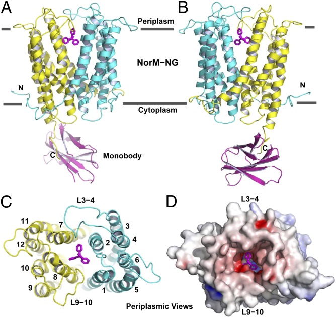
Fig. 1.
Structure of NorM-NG-monobody complex. (A and B) Structure of NorM-NG monobody complex as viewed from the membrane plane. The views in A and B are related by ∼180° rotation around the membrane normal. The amino (residues 5–230) and carboxyl (residues 231–459) halves of NorM-NG are colored cyan and yellow, respectively. Monobody is shown as a magenta ribbon and bound TPP as magenta sticks. (C) The arrangement of transmembrane helices in NorM-NG as viewed from the periplasmic side. (D) NorM-NG surface as viewed from the periplasmic side, which is colored according to electrostatic potentials from −20 (red) to +20 kTe−1 (blue).

