Fig. 7.
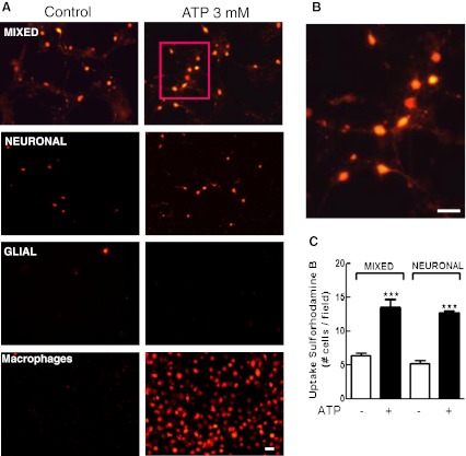
Uptake of sulforhodamine B (SRB) induced by ATP in the three types of retinal monolayer cultures. Mixed and neuronal retinal cultures at E7C2 or glial cultures were treated with 3 mM ATP for 10–15 min in the presence of 5 μM SRB in Hanks’ balanced salt solution without Ca++/Mg++. After incubation, cells were washed and visualized under fluorescence illumination. a Representative micrograph showing SRB labeled cells in mixed, purified neuronal, glial retinal cultures, and cultured macrophages. b High magnification of the cells shown in the red square in a. Note the presence of labeled neuronal processes. c Quantification of SRB-positive cells in mixed and neuronal retinal cultures. Labeled cells in each culture were counted in ten different micrographs randomly obtained. Data represent the mean ± SEM of five separate experiments performed in duplicate. ***p < 0.001, compared with control cultures. Bars = 20 μm

