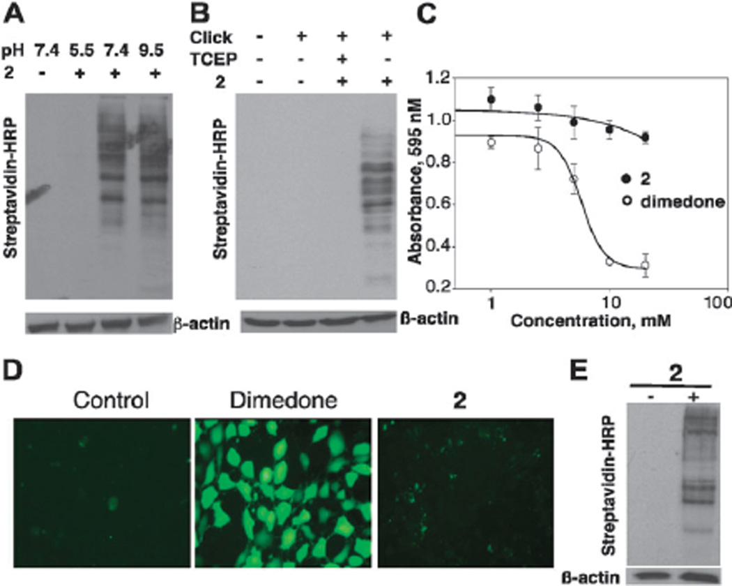
Fig. 3.
(A) Labeling of oxidized proteins with 2 at pH 5.5, 7.4 and 9.5 was monitored by Western blot. (B) Selectivity of −SOH labeling with 2 in lysates: cell lysates pre-reduced with TCEP were incubated with 2 at rt for 1 h (lane 3). (C) MTT assay to determine cell toxicity. (D) Intracellular ROS level was determined using DCF labeling in NIH 3T3 cells treated with 10 mM dimedone and 2. (E) Cell permeability assay. Cells treated with 2 (10 mM) for 2 h were washed extensively and then lysed in the absence of 2. Click reaction was performed to attach the biotin tag followed by Western blot analysis.

