Fig. 1.
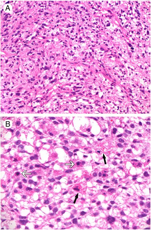
Histological features of UES. A, Short-spindled and ovoid tumor cells distributed in a loose stroma with delicate vasculature (original magnification, ×200). B, Intracytoplasmic hyaline globules noted in some tumors (black arrows). Note the presence of frequent mitotic figures (white arrows; original magnification, ×400).

